
Our Science
Conformational Epitope Mapping: Why the Linear vs Conformational Distinction Matters
- A linear epitope is a contiguous stretch of antigen sequence. A conformational epitope is assembled from residues that are distant in sequence and brought together by the fold.
- Most antibody-antigen interactions involve conformational epitopes.
- Conformational epitope mapping requires a method that reads the antibody-antigen complex with the antigen folded, in solution.
- CovalX maps both classes with high resolution by mass spectrometry.
Epitope mapping is the experimental identification of the binding site of an antibody on its target antigen. A linear epitope is formed by a continuous sequence of amino acids. A conformational epitope, also called a discontinuous epitope, is formed by amino acids that are close in the folded structure but distant in the sequence. Conformational epitope mapping therefore depends on methods that read the binding interface with the antigen in its native, folded state.
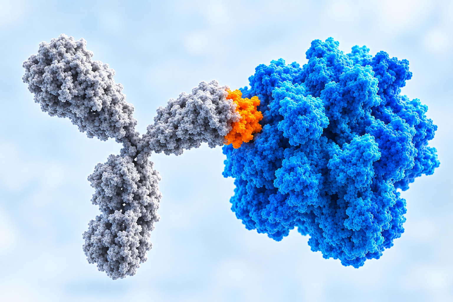 Epitope Mapping
What is an epitope?
An epitope is the part of an antigen that is recognized by the immune system. The part of the antibody that binds to the epitope is called a paratope. The epitopes of protein antigens can be conformational epitopes or linear epitopes, based on their structure and interaction with the paratope.
What is the difference between a linear and a conformational epitope?
A linear epitope is a binding site formed by contiguous residues of the antigen sequence, while a conformational epitope is a binding site formed by residues brought together by the three-dimensional folding of a protein.
| Linear epitope | Conformational epitope | |
|---|---|---|
| Sequence | One contiguous segment | Several discontinuous segments |
| Structural dependence | Less dependent on folding | Dependent on the local tertiary or quaternary structure |
| Natural occurrence | Less common (about 10% of B-cell epitopes) | Predominant form (about 90% of B-cell epitopes) |
| Effect of denaturation | Remains recognizable | Loses recognition |
| Common mapping approach | Overlapping peptide scanning, Western blot, immunohistochemistry | Native-state approaches, HDX-MS, XL-MS |
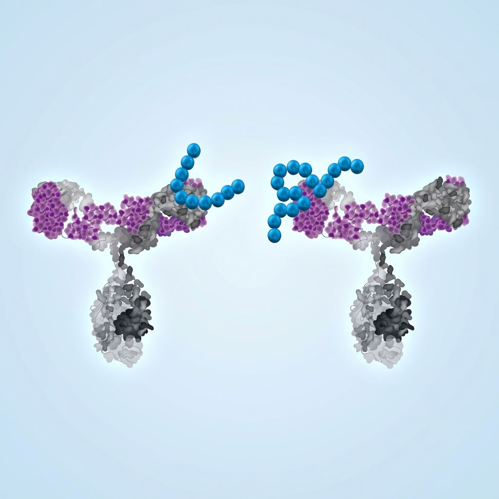 Conf vs Linear Epitope Mapping
Why the distinction between linear and conformational epitopes is a continuum
The linear/conformational split describes a gradient more than two separate categories. Even a linear epitope depends on more than its own residues: flanking residues and distant residues of the antigen affect whether that segment can adopt the conformation the antibody recognizes. Conformational epitopes, in turn, can include some linear segments.
Structural data put numbers on this gradient. Ferdous et al. analyzed the structures of 488 B-cell epitopes. Of these, 14% were made of a single region, and only 4% were strictly linear. In practice, “linear” usually designates an epitope dominated by one segment, whose binding still depends on local structure.
How does CovalX perform conformational epitope mapping?
CovalX maps linear and conformational epitopes with two mass spectrometry methods, Hydrogen Deuterium Exchange Mass Spectrometry (HDX-MS) and Cross-Linking Mass Spectrometry (XL-MS). Both analyses the antigen-antibody complex in solution, in its native state. Neither method requires the epitope class to be known in advance. The choice between them depends on the properties of the target, such as size, glycosylation, and disulfide bonds, and on the information the program needs.
WANT TO KNOW MORE?
Request a meeting with our team
Step 1: verifying the intact complex by High-Mass MALDI MS
Every CovalX epitope mapping project starts with High-Mass MALDI MS analysis of the intact antibody, the intact antigen, and the antibody-antigen complex. This step verifies:
- Antibody and antigen integrity,
- Antibody aggregation,
- Antigen multimerization,
- Stoichiometry of the complex.
For a conformational epitope, this is an important QC step. If the antigen is degraded, aggregated, or present in an unexpected conformational or oligomeric state, the epitope analysis may not be representative of the native state interaction.
HDX-MS: the epitope as protection from deuterium exchange
HDX-MS measures the rate at which backbone amide hydrogens of a protein exchange with deuterium from the solvent. That rate depends on solvent accessibility and hydrogen bonding. For epitope mapping, the free antigen and the antibody-antigen complex are incubated in D₂O in parallel. Regions that incorporate less deuterium in the presence of the antibody are the epitope candidates.
Protection reflects the three-dimensional binding interface. A conformational epitope therefore appears as several protected peptides from distant parts of the sequence, resolved in the same workflow as a linear epitope. HDX-MS also detects conformational changes induced by binding outside the contact surface, in the same dataset. Spatial resolution reaches one to five amino acids, depending on pepsin peptide coverage of the antigen.
XL-MS: the epitope as distance constraints
XL-MS uses a bifunctional cross-linker with a spacer arm of fixed length to covalently link residues in spatial proximity. The cross-linked peptides are then identified by tandem mass spectrometry (MS/MS). The result is a list of residue pairs across the antibody-antigen interface, expressed as distance constraints. This makes it suited to conformational epitopes.
Multiple enzymes are used in parallel to maximize sequence coverage. Epitope and paratope information are obtained from a single dataset.
HDX-MS or XL-MS : Which method for a conformational epitope?
Both HDX-MS and XL-MS can be used for conformational or linear epitope mapping. The properties of the target determine which method is best suited for a particular protein.
Find more information here : Epitope Mapping by Mass Spectrometry Epitope Mapping Overview
WANT TO KNOW MORE?
Request a meeting with our team
Conformational epitope mapping at CovalX
CovalX offers conformational and linear epitope mapping as part of its mass spectrometry service portfolio. The CovalX HDX-MS epitope mapping service and the CovalX XL-MS epitope mapping service pages detail each workflow, the deliverables, and the sample requirements. Every project includes High-Mass MALDI MS characterization of the intact complex.
Frequently asked questions
Can mass spectrometry map conformational epitopes?
Yes. HDX-MS identifies conformational epitopes by detecting the residues an antibody shields from solvent when it binds. XL-MS reports the residues at the interface through covalent distance constraints. Both methods work on folded proteins in solution.
Does conformational epitope mapping by mass spectrometry require crystallization?
No. HDX-MS and XL-MS both analyze the antigen and the antibody-antigen complex in solution under near-native conditions. This is a practical difference from X-ray crystallography, which requires a crystallized complex.
Does denaturation destroy a conformational epitope?
Denaturation disrupts the fold that brings the residues of a conformational epitope together, and binding is reduced or lost.
What determines the resolution of conformational epitope mapping?
In HDX-MS, resolution depends on the coverage of the antigen by overlapping peptides. In XL-MS, resolution depends on the position of reactive residues and on the sequence coverage obtained across the enzymes used. Both techniques reach one to five amino acids.
How can I tell if my antibody recognizes a conformational epitope?
Comparing binding to native and denatured antigen gives a first indication. If the antibody loses binding on a denaturing western blot but still binds in a native ELISA, it likely recognizes a conformational epitope. A residue-level assignment requires a structural method such as HDX-MS or XL-MS.
Can HDX-MS and XL-MS be combined to map the same conformational epitope?
Yes, HDX-MS and XL-MS can be combined to study the same epitope. The two datasets are independent, and the two methods can be used in orthogonal studies or to strengthen dossiers.