Our Science
Epitope Mapping by Mass Spectrometry: Locating Where an Antibody Binds Its Antigen
- Epitope mapping identifies the region of an antigen bound by an antibody. That surface shapes mechanism of action, developability, and the scope of related patent claims.
- Epitopes are either linear, a continuous stretch of sequence, or conformational, residues brought together by folding. Most antibody-antigen interactions involve conformational epitopes.
- Mass spectrometry maps epitopes in solution, under near-native conditions, across a broad range of target sizes and complex types.
- CovalX combines HDX-MS and XL-MS to map a wide range of epitope, antigen, binder, and selects the technique around each project.
Epitope mapping is the process of identifying the region of an antigen that an antibody, or another binder, recognizes. Where the antibody attaches to its antigen determines how it works: its mechanism of action, its potency, its risk of immunogenicity, and the scope of what a company can claim and protect. Mass spectrometry locates that region in solution, on the protein, for both linear and conformational epitopes.
Mass spectroscopy covers a wide range of targets, from small proteins to large complexes, as well as a wide range of binder format, from monoclonal antibodies, bispecifics, fragments, to non-antibody binders. Mass spectrometry maps all of them in solution, under near-native conditions.
This article explains what an epitope is, how mass spectrometry locates one, and why the right approach depends on your target rather than on a fixed method.
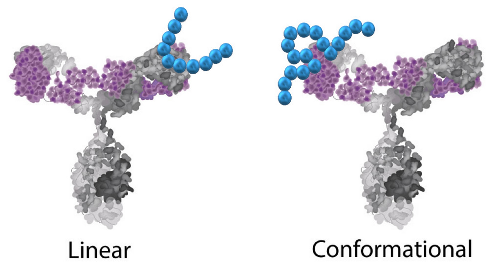 Coval-X linear conformational epitope mapping service
What is an epitope, and why does it matter for antibody drugs?
An epitope is the specific region of an antigen recognized by a binder, such as an antibody. Epitopes fall into two classes:
– A linear epitope is formed by a continuous stretch of amino acids in the sequence.
– A conformational epitope is formed by residues that are distant in the sequence but brought together when the protein folds into its three-dimensional structure.
Most antibody-antigen interactions involve conformational epitopes, which is why our mapping methods work on the folded, native protein rather than on sequence alone.
Mapping your epitope supports several development decisions. It clarifies mechanism of action, since the epitope determines which functional site of the target is engaged, and it separates candidates that hit the same region from those that reach distinct sites during lead selection and engineering. In comparability and biosimilar programs, epitope data provide evidence that a molecule engages its target the same way as a reference. A defined epitope also supports intellectual property, where it can ground or defend patent claims.
How does mass spectrometry map epitopes?
Mass spectrometry maps epitopes by analysing the antigen and its binder in solution, where the antigen keeps its native fold, so conformational epitopes are identified directly rather than inferred from sequence. CovalX uses two complementary methods. HDX-MS compares hydrogen-deuterium exchange between the free antigen and the complex. XL-MS detected covalent cross-links between residues that lie close across the interface, and the cross-linked peptides locate the contact sites directly.
The mass spectrometry epitope mapping toolkit
CovalX uses two mass spectrometry techniques for epitope mapping, chosen and combined according to the target.
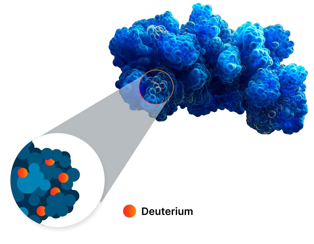 epitope_mapping_XL_MS Deuterieum exchange - XL-MS services, epitope mapping for linear, discontinuous and conformational epitope and paratope
HDX-MS: mapping by solvent protection
Hydrogen Deuterium Exchange Mass Spectrometry (HDX-MS) measures how readily the backbone amide hydrogens of a protein exchange with deuterium in solution, which reports on solvent accessibility and conformational dynamics. HDX-MS compares deuterium uptake by the antigen alone and in complex; the peptides that take up less deuterium in the presence of the binder mark the binding region. The method works in solution under near-native conditions and reports on conformational dynamics beyond the epitope itself. Spatial resolution depends on peptide coverage, which is a consideration on larger targets rather than a fixed size limit; for a large target, the workflow is adapted or paired with a second method for optimum resolution.
Related reading:
HDX-MS service page
Compound binding analysis by HDX-MS
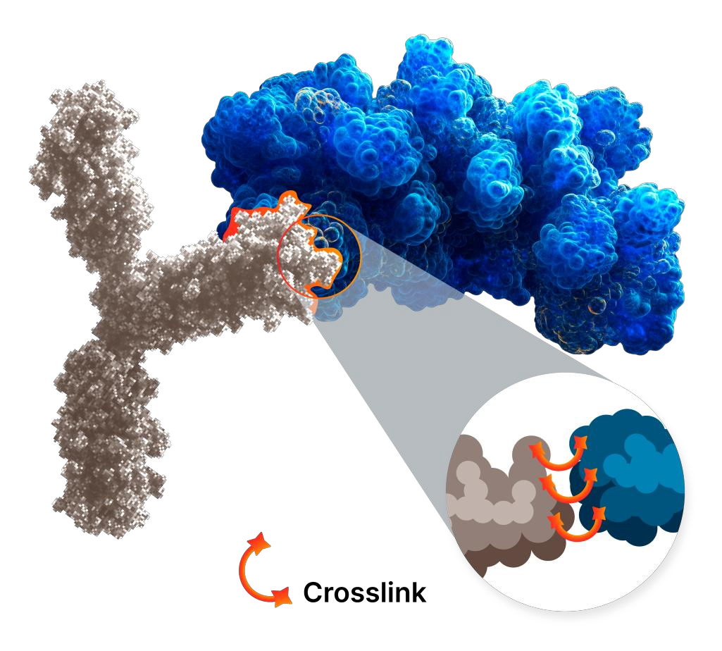 epitope_mapping_XL_MS Crosslinking zoom with XL-MS epitope mapping for linear, discontinuous and conformational epitope determination
XL-MS: mapping by distance constraints
Cross-Linking Mass Spectrometry (XL-MS) uses a bifunctional cross-linker in the form of a fixed spacer arm. The cross-linker creates a covalent bridge between residues within a defined distance, and tandem mass spectrometry identifies the linked peptide pairs. A bridge forms only between residues that were close at the moment of reaction, so the linked pairs report which parts of the antigen and binder sit at the interface. XL-MS handles large assemblies, glycosylated proteins, and structures stabilized by disulfide bonds, and it resolves interface geometry and overall architecture. It extends epitope mapping to targets where peptide-level protection is harder to read.
Related reading: XL-MS service page
High-Mass MALDI: Our QC step
Before epitope mapping starts, High-Mass MALDI mass spectrometry confirm the integrity of the interacting proteins and complexes. It measures the molecular weight of proteins and protein complexes up to 2 megadalton, using detection technology CovalX developed. The measurement resolves the stoichiometry of the complex: how many proteins of each partner are bound, and at what total mass. It confirms the assembly is intact and at the expected mass, and detects aggregates and multimerization. It measures each of these directly, at the mass accuracy of the spectrometer.
Epitope mapping follows only once the proteins and complexes pass this check. Mapping a validated complex is one reason CovalX analyses reach a high success rate. A map built on a sample that does not represent the intended biology returns binding sites for an artefactual interaction.
Related reading: High-Mass MALDI service page
What kinds of epitopes and targets can mass spectrometry map?
Mass spectrometry epitope mapping applies across epitope types, target sizes, and binder formats. Three variables define the range:
- Epitope type. Linear and conformational epitopes are both mapped on the folded protein in solution, so discontinuous surfaces are read directly.
- Target size and complexity. Targets from small single-domain proteins to large complexes, glycosylated, or disulfide-stabilized assemblies fall within range.
- Binder format. Monoclonal antibodies, bispecifics, antibody fragments, chemical compounds, peptides and non-antibody binders are all compatible, since the readout depends on the interface rather than on the binder’s class.
Target characteristics guide method selection at CovalX, rather than the target being fitted to a single available method.
WANT TO KNOW MORE?
Request a meeting with our team
Work with CovalX
The right approach depends on the epitope, the target, and the binder in front of you. CovalX runs these techniques across that range and selects, or combines, them around your case. CovalX provides epitope mapping through its HDX-MS and XL-MS epitope mapping service, with High-Mass MALDI control to validate the intact complex beforehand. For programs that also need subunit analysis of the binder, the CovalX Antibody characterization service covers those measurements.
Discuss your epitope mapping project with our team, and we will propose an approach built for your target.
Frequently asked questions
What can mass spectrometry epitope mapping map?
Mass spectrometry maps both linear and conformational epitopes on the folded protein in solution. It applies to targets from small proteins to megadalton assemblies, including glycosylated and disulfide-stabilized proteins.
Does target size limit mass spectrometry epitope mapping?
Target size influences which technique fits best, not whether mapping is possible. HDX-MS resolution depends on peptide coverage, which is a consideration on very large targets, while XL-MS is suited medium size target 10-100 kDa to large assemblies and complexes. At CovalX, every project is discussed, and for large or complex targets, the workflow is adapted.
How does HDX-MS locate an epitope?
HDX-MS compares how readily backbone amide hydrogens exchange with deuterium for the antigen alone and in complex. Residues shielded at the interface exchange more slowly, so peptides with reduced deuterium uptake in the complex mark the binding region.
How does XL-MS locate an epitope?
XL-MS uses a chemical cross-linker with a fixed spacer arm to covalently bridge residue pairs that sit within a defined distance in the antibody-antigen complex. Cross-links that span the interface are identified by MS after digestion, so the antigen residues linked to the antibody mark the binding region within the spacer arm’s distance limit.
Can mass spectrometric epitope mapping data be used in regulatory submissions?
Mass spectrometry structural characterization can be included in IND and BLA packages as structural evidence. Both the FDA and the EMA accept MS-based structural characterization data.
Can a single study use more than one mass spectrometry method?
Yes. HDX-MS and XL-MS are complementary, orthogonal methods, and are often combined in one project.